- HomePage
- News
- Research Interests
- Methods
- Publications
- Software
- Teaching
- Group Members
- Contact
- Photos
For student and postdoctoral enquiries (with CV), e-mail
Professor Achilles Kapanidis.
Funding sources:


Single-molecule fluorescence spectroscopy
We apply single-molecule fluorescence spectroscopy (SMFS) to study biomolecular interactions. We exploit fluorescence resonance energy transfer (FRET) at the single-molecule level, combined with alternating-laser excitation (ALEX) spectroscopy (see references). Our experiments are carried out both in solution as well as on a surface, using total internal reflection fluorescence (TIRF) microscopy.
We currently operate two single-molecule setups in our spectroscopy laboratory: a three-colour ALEX setup (473 nm, 532 nm and 635 nm) and a combined two-colour ALEX / TIRF setup (532 nm, 635 nm).
A. 3-colour ALEX (3c-ALEX) setup
We extended the principle of ALEX spectroscopy in solution by adding a third excitation wavelength, and currently operate three lasers with wavelengths of 473, 532 and 635 nm at out 3c-ALEX setup.
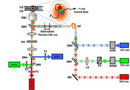
3-color ALEX setup
Legend:
-AOM: Acousto-Optical Modulator
-EOM: Electro-Optical Modulator
-DM: Dichroic Mirror
-P: Polariser
-λ/4: Quarter-Wavelength Plate
-MR: Mirror
-OF: Optical Fibre
-OBJ: Objective
-CS: Coverslip
-PH: Pinhole
-F: Filter
-APD: Avalanche Photo-Diode
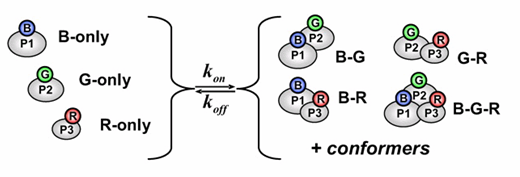
3c-ALEX probes three different fluorophores, and thus three distances (using FRET) and three stoichiometries become accessible. The additional information makes 3c-ALEX an ideal tool to study biomolecular interactions, which involve interplay of many molecules, such as proteins, DNA or RNA.
Further information:
-Nam Ki Lee, Achillefs N. Kapanidis, Hye Ran Koh, You Korlann, Sam On Ho, Younggyu Kim, Natalie Gassman, Seong Keun Kim, and Shimon Weiss; Biophys. J., 2007, 92, 303-312. Three-Color Alternating-Laser Excitation of Single Molecules: Monitoring Multiple Interactions and Distances pubmed citation
-Joachim Ross, Peter Buschkamp, Daniel Fetting, Achim Donnermeyer, Christian M. Roth, and Philip Tinnefeld; J Phys Chem B, 2007, 111(2), 321-326. Multicolor single-molecule spectroscopy with alternating laser excitation for the investigation of interactions and dynamics
pubmed citation
B. 2-colour ALEX / TIRF setup
We built a two-colour ALEX setup 532 and 635 nm) which allows both confocal SMFS in solution, as well as TIRF microscopy on a surface.
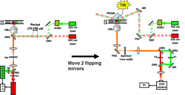
2-color ALEX / TIRF setup
The combination of the ALEX principle with TIRF microscopy is realized by spectrally separating the fluorescence emission. The emitted photons, collected through a water immersion objective, are split into two regions of the CCD camera using two 630DRLP dichroic mirrors (dual-view format), allowing for simultaneous observation of the donor and acceptor fluorescence. The synchronization of the alternation with the integration periods of the camera is achieved using the frame readout output signal of the camera to trigger the alternation of the laser sources.
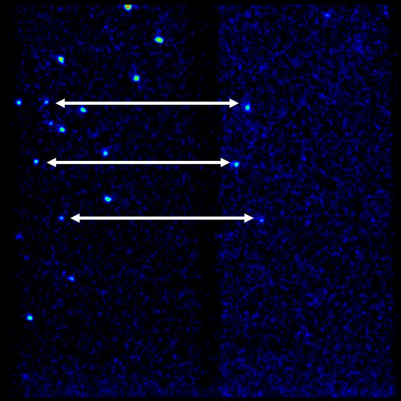
TIRF image of a RNA polymerase-DNA complex. The DNA is doubly labeled with a green fluorophore (Cy3B) and a red fluorophore (ATTO647N). The green emission channel is on the left, the red emission channel on the right side of the field of view of the CCD image. This snapshot shows Green excitation only, the spots on the red channel show FRET
For further information:
-Emmanuel Margeat, Achillefs N. Kapanidis, Philip Tinnefeld, You Wang, Jayanya Mukhopadhyay, Richard H. Ebright, Simon Weiss; Biophys J, 2006, 90(4), 1419-1431. Direct observation of abortive initiation and promoter escape within single immobilized transcription complexes. Pubmed citation