|
|
GALLERY OF MOVIES
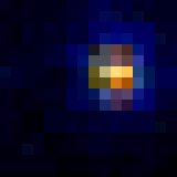
Digital Holographic microscopy:
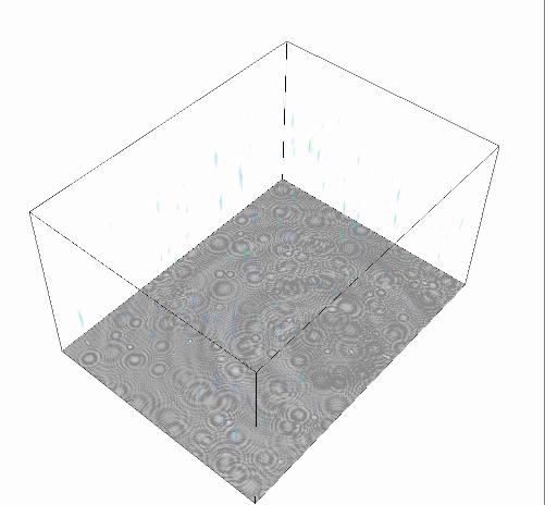
Inline holographic movie and 3D reconstruction of a population of swimming
E. coli. |
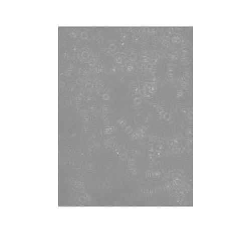
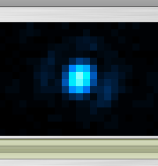
Digital re-focussing through the 3D volume reconstructed from one
frame of an in-line holographic movie of a population of swimming
E. coli. 70 microns focal depth in 0.5 micron steps. The movie
starts in the plane of the hologram. |
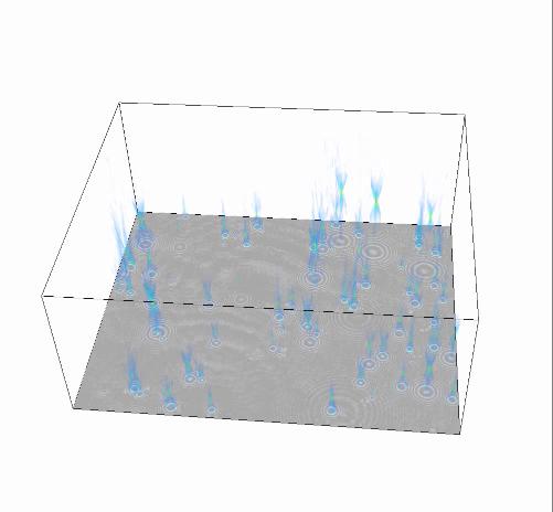
Inline holographic movie and 3D reconstruction of Brownian Motion of
polystyrene beads. |
Bacterial Flagellar motor:
1 micron beads spinning on E. coli flagellar motors, real speed. |
A 200 nm fluorescent bead spinning with 26 steps per revolution on a
sodium-driven chimeric flagellar motor. 30x slower than real speed,
video recorded at 2400 frames per second. |
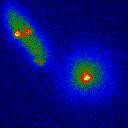
A 100 nm gold bead spinning at 900 Hz on a sodium-driven chimeric
flagellar motor. 2000x slower than real speed, video recorded at 100,000
frames per second using laserdark-field microscopy. |
E. coli cells expressing GFP-labelled motor protein MotB. The
same two cells are shown in fluorescence (left, slowed x2) and
bright-filed (right, real speed). Photobleaching of the motor allows
counting of the number of MotB molecules |
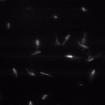
Swimming E. coli with fluorescently labelled flagella and
bundle formation disrupted by streptavidin binding to biotin-labelled
flagellar hooks. |
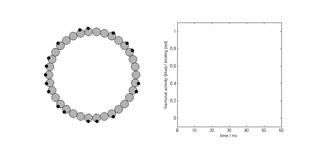
Simulation of conformational spread in the Flagellar rotor, as a
mechanism for co-ordinated switching of the motor. |
F1-ATPase

A 60 nm gold bead attached to an F1-ATPase molecule from
Yeast, spinning with 3 steps per rev, viewed with laser darkfield
microscopy. Slowed down 200x. |
 
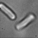
A pair and a triplet of 500 nm polystyrene beads attached to an F1-ATPase
molecule from E. coli, viewed with bright-field microscopy.
Real speed. |
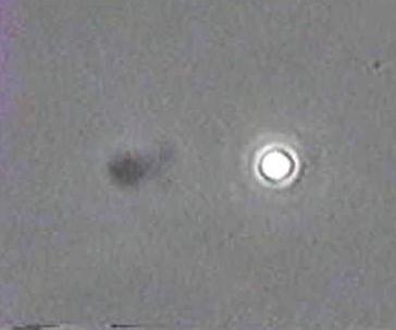
Optical traps and Electrorotation:
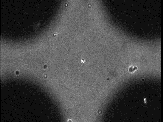 4.8 MB .avi (Higher magnification: 2.3 MB AVI) Electrorotation of a 1 micron polystyrene bead, decorated with 0.2
micron beads, held in an optical trap. |
|
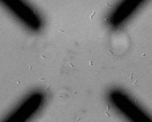 1.6 MB .mov (Higher magnification: 1.7 MB .mov) Electrorotation of a tethered E. Coli cell.
|
last updated: 11 September 2012